Monday, September 30, 2013

Researchers Demonstrate 'Accelerator on a Chip'

Menlo Park, Calif. — In an advance that could dramatically shrink particle accelerators for science and medicine, researchers used a laser to accelerate electrons at a rate 10 times higher than conventional technology in a nanostructured glass chip smaller than a grain of rice.
The achievement was reported today in Nature by a team including scientists from the U.S. Department of Energy’s (DOE) SLAC National Accelerator Laboratory and Stanford University.
“We still have a number of challenges before this technology becomes practical for real-world use, but eventually it would substantially reduce the size and cost of future high-energy particle colliders for exploring the world of fundamental particles and forces,” said Joel England, the SLAC physicist who led the experiments. “It could also help enable compact accelerators and X-ray devices for security scanning, medical therapy and imaging, and research in biology and materials science.”
Because it employs commercial lasers and low-cost, mass-production techniques, the researchers believe it will set the stage for new generations of "tabletop" accelerators.
At its full potential, the new “accelerator on a chip” could match the accelerating power of SLAC’s 2-mile-long linear accelerator in just 100 feet, and deliver a million more electron pulses per second.
This initial demonstration achieved an acceleration gradient, or amount of energy gained per length, of 300 million electronvolts per meter. That's roughly 10 times the acceleration provided by the current SLAC linear accelerator. 
“Our ultimate goal for this structure is 1 billion electronvolts per meter, and we’re already one-third of the way in our first experiment,” said Stanford Professor Robert Byer, the principal investigator for this research.
This animation explains how the accelerator on a chip uses infrared laser light to accelerate electrons to increasingly higher energies. (Greg Stewart/SLAC)

How It Works

Today’s accelerators use microwaves to boost the energy of electrons. Researchers have been looking for more economical alternatives, and this new technique, which uses ultrafast lasers to drive the accelerator, is a leading candidate.
Particles are generally accelerated in two stages. First they are boosted to nearly the speed of light. Then any additional acceleration increases their energy, but not their speed; this is the challenging part.
In the accelerator-on-a-chip experiments, electrons are first accelerated to near light-speed in a conventional accelerator. Then they are focused into a tiny, half-micron-high channel within a fused silica glass chip just half a millimeter long. The channel had been patterned with precisely spaced nanoscale ridges. Infrared laser light shining on the pattern generates electrical fields that interact with the electrons in the channel to boost their energy. (See the accompanying animation for more detail.)
Turning the accelerator on a chip into a full-fledged tabletop accelerator will require a more compact way to get the electrons up to speed before they enter the device.  
A collaborating research group in Germany, led by Peter Hommelhoff at Friedrich Alexander Universityand the Max Planck Institute of Quantum Optics, has been looking for such a solution. Itsimultaneously reports in Physical Review Letters its success in using a laser to accelerate lower-energy electrons.

Multi-Use Accelerators

Applications for these new particle accelerators would go well beyond particle physics research. Byer said laser accelerators could drive compact X-ray free-electron lasers, comparable to SLAC’s Linac Coherent Light Source, that are all-purpose tools for a wide range of research.
Another possible application is small, portable X-ray sources to improve medical care for people injured in combat, as well as provide more affordable medical imaging for hospitals and laboratories. That’s one of the goals of the Defense Advanced Research Projects Agency’s (DARPA) Advanced X-Ray Integrated Sources (AXiS) program, which partially funded this research. Primary funding for this research is from the DOE’s Office of Science.
SLAC's Joel England explains how the same fabrication techniques used for silicon computer microchips allowed their team to create the new laser-driven particle accelerator chips. (SLAC)
The study's lead authors were Stanford graduate students Edgar Peralta and Ken Soong. Peralta created the patterned fused silica chips in the Stanford Nanofabrication Facility. Soong implemented the high-precision laser optics for the experiment at SLAC’s Next Linear Collider Test Accelerator. Additional contributors included researchers from the University of California-Los Angeles and Tech-X Corp. in Boulder, Colo.
SLAC is a multi-program laboratory exploring frontier questions in photon science, astrophysics, particle physics and accelerator research. Located in Menlo Park, California, SLAC is operated by Stanford University for the U.S. Department of Energy Office of Science. To learn more, please visitwww.slac.stanford.edu.
DOE’s Office of Science is the single largest supporter of basic research in the physical sciences in the United States, and is working to address some of the most pressing challenges of our time. For more information, please visit science.energy.gov.

Citation: E. A. Peralta et al.Nature, 27 Sept 2013 (10.1038/nature12664)

Press Office Contact: Andy Freeberg, SLAC, afreeberg@slac.stanford.edu, (650) 926-4359

Scientist Contacts:
Robert Byer, Stanford University, rlbyer@stanford.edu, (650) 723-0226
Joel England, SLAC, england@slac.stanford.edu, (650) 926-3706

*This post was taken from http://www6.slac.stanford.edu/news/2013-09-27-accelerator-on-a-chip.aspx* 
Dental implants that heal faster and fight infection


[+]
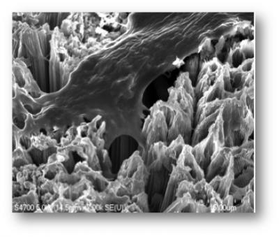
A bone cell anchoring itself to a surface of titanium dioxide nanotubes. Because osteoblasts (cells that synthesize bone tissue).readily adhere to this novel surface, dental implants coated with TiO2 nanotubes could significantly improve healing following dental implant surgery. (Credit: Tolou Shokuhfar/Michigan Technological University)
Michigan Technological University researchers have developed a way to use self-assembled titanium dioxide (TiO2) nanotubes to lower the rate of dental-implant failures.
Dental implants are posts, usually made of titanium, that are surgically placed into the jawbone and topped with artificial teeth.
While most dental implants are successful, a small percentage fail and either fall out or must be removed.
“There are two main issues that concern dentists: infection and separation from the bone,” said Tolou Shokuhfar, an assistant professor of mechanical engineering.
The mouth is a dirty place, so bacterial infections are a risk after implant surgery, and sometimes bone fails to heal securely around the device.
Because jawbones are somewhat thin and delicate, replacing a failed implant can be difficult, not to mention expensive. Generally, dentists charge between $2,000 and $4,000 to install a single implant, and the procedure is rarely covered by insurance.
[+]
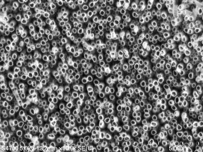
A forest of titanium dioxide nanotubes etched into metallic titanium. The surface may improve the longevity of dental implants. (Credit: Tolou Shokuhfar/Michigan Technological University)
Shokuhfar is now working with Cortino Sukotjo, a clinical assistant professor at the University of Illinois at Chicago (UIC) College of Dentistry on a dental implant with a surface made from TiO2 nanotubes, but she has been making and testing them for several years.
“We have done toxicity tests on the nanotubes, and not only did they not kill cells, they encouraged growth,” she said.
She has already demonstrated that bone cells grow more vigorously and adhere better to titanium coated with TiO2 nanotubes than to conventional titanium surfaces. That could keep more dental implants in place.
Drug delivery
The nanotubes can also be a drug delivery system. Shokuhfar’s team, in collaboration with Alexander Yarin, a professor in UIC’s Department of Mechanical and Industrial Engineering, loaded TiO2 nanotubes with the anti-inflammatory drug sodium naproxen and demonstrated that it could be released gradually after implant surgery.
That assures that the medicine gets where it’s needed, and it reduces the chances of unpleasant side effects that arise when a drug is injected or taken orally.
[+]
This cutaway view of a titanium dioxide nanotube reveals the drug sodium naproxen on the inside. On the surface of a dental implant, these nanotubes could release this anti-inflammatory drug or other pharmaceuticals that could speed healing. (Credit: Tolou Shokuhfar/Michigan Technological University)
To fight infection, the TiO2 nanotubes can also be laced with silver nanoparticles. Shokuhfar and Craig Friedrich, who holds the Richard and Bonnie Robbins Chair of Sustainable Design and Manufacturing at Michigan Tech, are conducting research, as yet unpublished, that is focused on orthopedic implants, such as artificial hips, but which also applies to dental implants.
“Silver has antimicrobial properties, and we are capable of obtaining a dose that can kill microbes but would not hurt healthy cells and tissues,” she said. In particular, it can help prevent biofilms, vast colonies of bacteria that can cover implants and be very difficult to eradicate. A nanotextured implant surface embedded with silver nanoparticles could prevent infection for the life of the implant.
The TiO2 nanotubes also have a cosmetic advantage: transparency. That’s a plus for any dental implant, but especially for a new type made from zirconia, which some patients choose because it is totally white.
Shokuhfar expects that implants with the new nanotubular surface will be easily assimilated into the market, since titanium implants, both dental and orthopedic, have a long history.
Shokuhfar and Friedrich have received a provisional patent and are working with two hospitals to further develop the technology and eventually license it. “As soon as the related paper work is taken care of and we get the FDA approval, the technology could be applied. However I am not aware how long all that would take,” she told KurzweilAI.
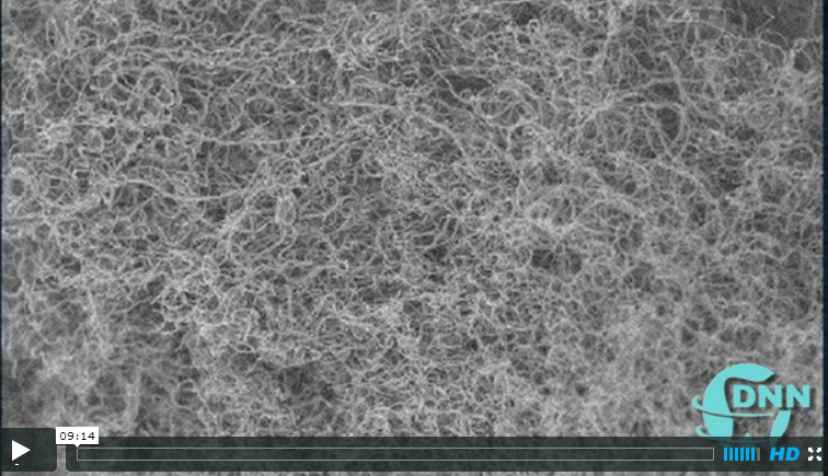
Interview with Dr. Tolou Shokuhfar (credit: DNN)

REFERENCES:

*All credit for this post goes to: http://www.kurzweilai.net/dental-implants-that-heal-faster-and-fight-infection * 

Wednesday, September 4, 2013

HIV Vaccine

“HIV/AIDS has killed 35 million people worldwide, and more than 34 million people currently live with the virus infection. Since the virus was characterized in 1983, there have been numerous trials through pharmaceutical companies and academic institutions around the world to develop vaccines; however, no vaccine has been successful to date.”

Researchers from Western University in Canada and Sumagen Canada Inc have successfully completed Phase I Clinical Trial of (SAV CT 01) the first and only preventative HIV vaccine that is based on a genetically modified killed whole virus (SAV001-H) has shown to be successful in all patients with no adverse side-effects.

The vaccine is a prophylactic vaccine, meaning that is not a cure for people who are already living with the HIV/AIDS virus; but a preventative treatment for individuals that have not been infected by the HIV virus. In an interview, Dr. Chil-Yong Kang explains that the vaccine could be beneficial in suppressing the virus in people who are already have HIV or with hepatitis C.


“Other HIV vaccines evaluated through human clinical trials have focused on either one specific component of HIV as an antigen, genetic vaccine using recombinant DNA, or recombinant viruses carrying the HIV genes. Kang’s vaccine is unique in that it uses a killed whole HIV-1, much like the killed whole virus vaccines for polio, influenza, rabies and hepatitis A. The HIV-1 is genetically engineered so it is safer and can be produced in large quantities.”

Scientist are optimistic about the results from the Phase I trials, because the vaccine boosted the antibody production in HIV-positive volunteers, which raises suspicion that Phase 2 human clinical trials will yield a substantial increased immune systems response to the HIV virus.

"We have proven that there is no safety concern of SAV001-H in human administration and we are now prepared to take the next steps towards Phase II and Phase III clinical trials," said Dr. Dong Joon Kim in the official release. "We are delighted to be one step closer to the first commercialized HIV vaccine."

“Sumagen anticipates not only having the first HIV vaccine in market but also the eradication of HIV/AIDS for human beings.”

Source: http://communications.uwo.ca/media/releases/2013/September/no_adverse_effects_in_volunteers_following_phase_i_clinical_trial_of_sumagen_aids_vaccine.html